A Step by Step Guide: On Animal’s Vaccination and Livestock
Animal Vaccination and Disease Prevention
Importance and Benefits of an Animal Vaccination:
What is Animal Vaccination?
Animal vaccination involves the administration of a vaccine, a biological substance designed to stimulate the immune system to develop immunity to a specific disease without causing the disease itself. Vaccines typically contain either killed or weakened forms of pathogens (bacteria, viruses), or components of these pathogens (like proteins), which allow the immune system to recognize and fight the actual pathogen in the future.
Vaccination is one of the most effective ways to prevent the spread of infectious diseases in animals, ensuring the health and well-being of individual animals and the broader community.
Why is Animal Vaccination Important?
- Prevention of Disease: Vaccination helps prevent the onset of serious and often fatal diseases. Some diseases, like rabies or distemper, can be deadly if not prevented by vaccination.
- Controlling Outbreaks: Vaccines help prevent the spread of contagious diseases, especially in animals living in close quarters (e.g., shelters, farms, zoos, and kennels).
- Public Health: Certain animal diseases, such as rabies, can also be transmitted to humans (zoonotic diseases). Vaccinating pets and livestock can prevent the transmission of these diseases to humans.
- Improved Productivity: In the case of livestock, vaccination improves productivity. Healthy animals are more likely to grow efficiently, produce milk, and reproduce, which benefits the farming industry.
- Long-Term Protection: Regular vaccination helps animals maintain immunity against diseases, ensuring they remain healthy throughout their lives.
Types of Vaccines for Animals
Vaccines can be divided into two main categories: Core and Non-core vaccines.
1. Core Vaccines
Core vaccines are considered essential for all animals in a particular species because the diseases they prevent are widespread, severe, and pose significant risks to animal health. This vaccines are usually recommended for all animals, regardless of their environment or lifestyle.

- For Dogs:
- Rabies: A fatal disease affecting the brain, caused by a virus that can also affect humans. Rabies vaccination is often required by law in many regions.
- Distemper: A contagious and potentially fatal viral disease that affects the respiratory, gastrointestinal, and nervous systems.
- Parvovirus: A severe viral disease that affects a dog’s gastrointestinal system, leading to vomiting, diarrhea, and often death.
- Adenovirus (Hepatitis): A viral disease that affects the liver and kidneys, potentially causing fatal damage.
- Parainfluenza: A viral infection that causes respiratory problems.
- For Cats:
- Rabies: As with dogs, rabies vaccination is essential for cats to prevent the disease’s spread.
- Feline Distemper (Panleukopenia): A potentially fatal viral infection affecting the gastrointestinal, immune, and nervous systems.
- Feline Herpesvirus: Causes respiratory illness and eye infections in cats.
- Feline Calicivirus: A virus that causes respiratory issues, mouth ulcers, and can lead to serious complications.
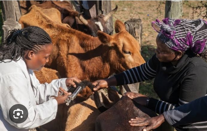
- For Livestock:
- Foot and Mouth Disease (FMD): A highly contagious viral disease that affects cloven-hoofed animals like cows, sheep, and goats.
- Brucellosis: A bacterial infection that affects reproductive organs in livestock, often leading to infertility.
- Bovine Tuberculosis: A bacterial infection affecting cattle, capable of being transmitted to humans (zoonotic).
- Avian Influenza: A viral infection affecting poultry, which can result in large-scale losses in the poultry industry.

2. Non-Core Vaccines
Non-core vaccines are recommended based on an animal’s specific needs, risks, and lifestyle. These vaccines might not be necessary for every animal, but they are given to protect animals in specific environments or situations.
- For Dogs:
- Leptospirosis: A bacterial infection transmitted through water or soil contaminated by the urine of infected animals. It can cause kidney and liver failure.
- Bordetella bronchiseptica: A bacterial infection that causes kennel cough, a highly contagious respiratory disease in dogs, especially in boarding kennels or dog parks.
- Lyme Disease: Caused by ticks, this disease can result in joint inflammation and other health problems in dogs.
- For Cats:
- Feline Leukemia Virus (FeLV): A viral infection that weakens the immune system and can lead to cancer in cats.
- Feline Immunodeficiency Virus (FIV): Known as cat AIDS, this virus weakens the immune system, making cats more susceptible to other infections.
- For Livestock:
- Clostridial diseases: Caused by bacteria in the soil, leading to fatal infections like tetanus and blackleg. Vaccines are the one that we usually give to livestock, especially cattle, to prevent these diseases.
- Peste des Petits Ruminants (PPR): A viral disease that affects goats and sheep, leading to high mortality rates, particularly in regions where these animals are essential for food security.
- Animal vaccination offers numerous benefits that contribute to the overall health and well-being of animals, as well as the people and environments they interact with. Here are the key benefits:
Vaccination Schedule
The timing of vaccines depends on the species, age of the animal, and the specific disease we will be vaccinating to.
- Puppies and Kittens: Vaccinations usually begin at 6-8 weeks of age, with booster shots every 3-4 weeks until they reach about 16 weeks old.
- Adult Animals: After the initial vaccination series, adults may require booster shots every 1-3 years, depending on the vaccine and the animal’s risk factors.
- Livestock: Livestock often receive vaccinations based on their age, the type of animals, and regional disease risks. We make sure we give some vaccine annually, while others may require more frequent doses.
- Most places likev SPCA will not want to vaccinate your pet if you are not going to sterilize also.
Also Read About :Animal External Parasites
Learn More About: Bluetongue disease in Sheeps
Vaccine Safety
Vaccines are typically safe, and side effects are rare. The most common side effects are mild, such as soreness at the injection site, mild fever, or fatigue. In rare cases, animals may experience allergic reactions, but these are typically treatable with immediate veterinary care.
It’s important to follow the recommended vaccination schedule and consult a veterinarian for the appropriate vaccines based on the animal’s age, environment, and potential exposure risks.
Find Other Vaccination Campaigns
Conclusion
Animal vaccination is an essential part of responsible pet ownership and livestock management. It protects animals from a wide range of dangerous diseases, ensures the health and safety of both the animals and humans around them, and contributes to disease control on a broader scale. By working with a veterinarian, pet owners and livestock farmers can create an effective vaccination plan to maintain healthy animals and prevent the spread of infections.


